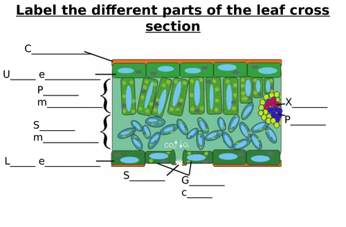
Tradescantia Leaf Cross Section Labeled

Anatomy Of Tradescantia Fluminensis A B Cross Section Of Stem C F Download Scientific Diagram
Biolympiads Com Wp Content Uploads 18 09 Plant Structure And Development Pdf

70 Plant Anatomy Ideas Microscopic Photography Patterns In Nature Microscopic

Unbiol1

Plant Structure Growth And Development Ppt Download
Organography And Plant Histology Of Rhoeo Discolor Tradescantia Spathacea Steemit
Start studying Leaf Cross Section Learn vocabulary, terms, and more with flashcards, games, and other study tools.

Tradescantia leaf cross section labeled. Crosssection of a deciduous leaf (Item No P) Curricular Relevance Additional Requirements Blatt einer zartblättrigen Pflanze, zB Tradescantia Holundermark oder Rübe Water Ethanol Experiment Variations Keywords Task and equipment Information for teachers Information The leaf of a deciduous plant consists of several layers. (question 6 and 9) 1 Everyone should read and complete the lab. GEOPAK Road 1 Chapter 9 – Cross Sections & Labeling 4/12/16 Missouri Department of Transportation 95 Spacing Leaf Use this dialog to define the settings for stacked or sheet modes section) Distance between sections.
Anthemweus is your first and best source for all of the information you’re looking for From general topics to more of what you would expect to find here, anthemweus has it all We hope you find what you are searching for!. Hymenachne (marsh grass) freehand cross section of leaf with bulliform cells, aerenchyma, and clusters of sclerenchyma Acetate peel from upper epidermis of bean ( Phaseolus vulgarus ) leaf Tradescantia (spiderwort) upper epidermis. (a) Draw the diagram of cross section of a leaf and label the following parts (i) chloroplast (ii) cuticle (b) A gas is released during photosynthesis Name the gas and also state the way in which the gas is evolved (c) In certain group of plants, stomata remains closed during day How is food synthesized by such plants Also name them.
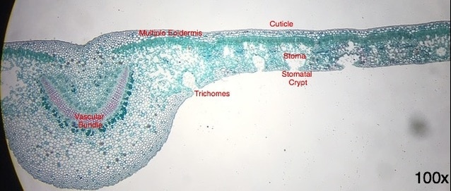
Leaf blade crosssection the leaf blade is more or less flat in crosssection 2×Tradescantia subaspera KerGawl is a very rare spiderwort hybrid likely originating from a garden planting It is known from MA It has glandular hairs on the sepals. Ficus (fig) leaf cross section with multiple epidermis layers and lithocyst in the upper epidermis Ficus (fig) leaf cross section Oleander leaf cross section with stomatal crypts, thick cuticle, and multiseriate epidermis. Apical Meristems Primary and Secondary Growth Leaf Anatomy Fall 11 Cell Types From Berg L 1997 Introductory Botany Saunders College Publishers Stem (Tradescantia) leaf (LM) Epidermal cell (b) Fig 3618 Ocotillo (leafless) Oleander leaf cross section and flowers Cuticle Upper epidermal tissue Ocotillo leaves Trichomes (“hairs.
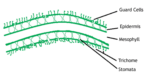
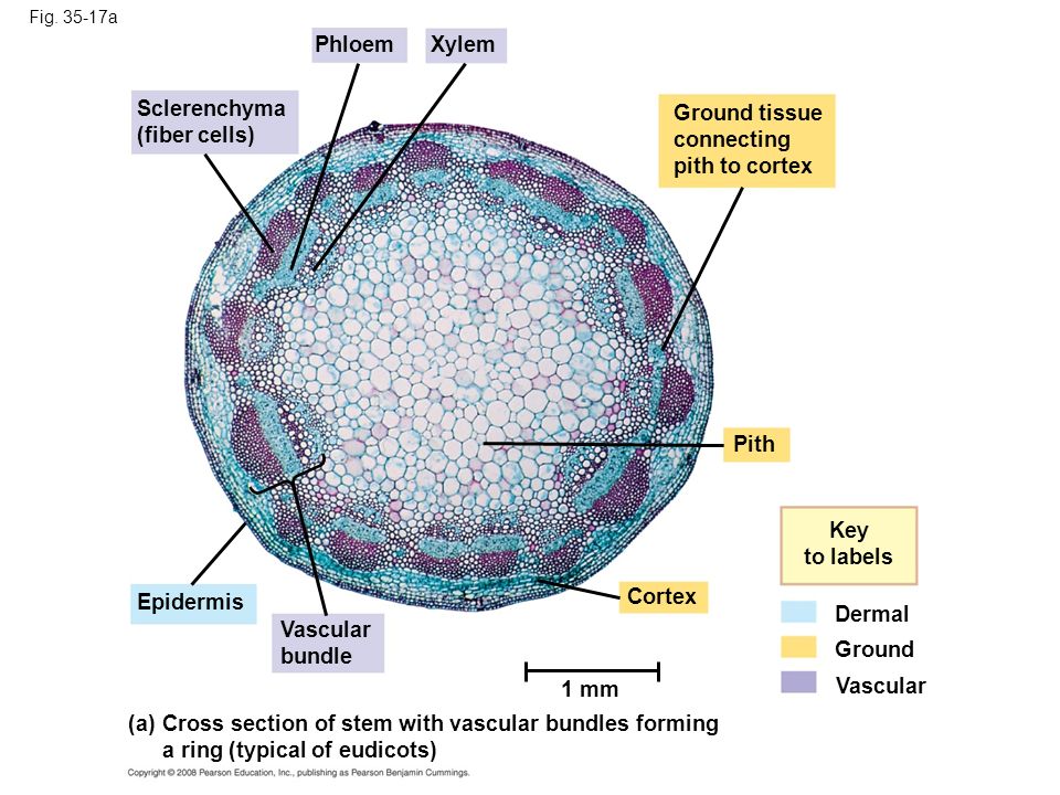
(Tradescantia) leaf (LM) Epidermal cell (b) m Fig 3518c Upper epidermis Palisade mesophyll Key to labels Dermal Ground Vascular Spongy mesophyll Lower epidermis Vein Air spaces Guard cells Cross section of a lilac (Syringa) leaf (LM) (c) µm Secondary Growth •Secondary growth adds girth to stems and roots in woody plants, (it doesn’t. Name the product produced on the leaf cross section label #1 Preview this quiz on Quizizz In plants, what is one of the draw backs (negative side effects) of releasing oxygen gas and taking in carbon dioxide?. Balsa Wood (cross section) Showing Large Conductive Elements (SEM x2) This image is copyright Dennis Kunkel at wwwDennisKunkelcom , used with permission Details of the stem of basswood.
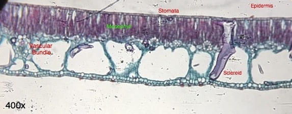
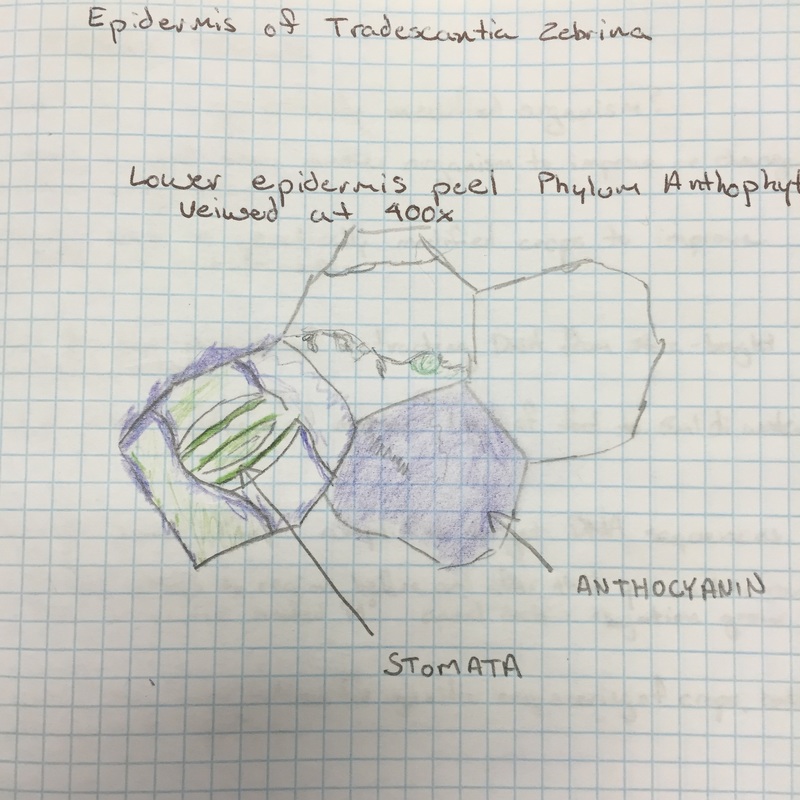
This is a cross section through a rice leaf blade Click here for a labeled picture Dermal Tissue System Epidermis Stomata Bulliform Cells Vascular Tissue System Xylem Phloem Bundle Sheath Fiber Ground Tissue System Mesophyll Aeranchyma Branched Parencyma. Tradescantia zebrina (spiderwort) Perform a cross and a paradermal section Perform a cross and a paradermal section The simplest case we could imagine for an epidermis would be like the upper (adaxial) epidermis on the Tradescantia leaf. This is a thumbnail of the leaf cross section Label Me!.
Large surface area Maximise light absorption Thin Short distance for carbon dioxide to diffuse into leaf. Oct 12, 18 This website is for sale!. Microphotographs Of The Leaf Cross Section Under Light Microscopy A Scientific Diagram Tradescantia Leaf Stomata Prepared Microscope Slide China 50pcs Set For Botany Leaf Microscope Prepared Slides Iris Leaf Epidermis Prepared Microscope Slide Basic Anatomy.
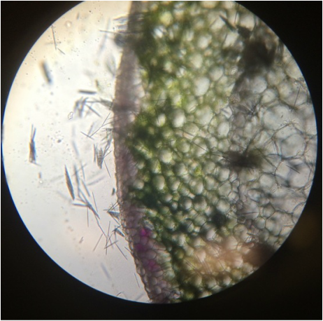
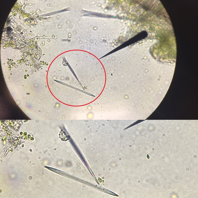
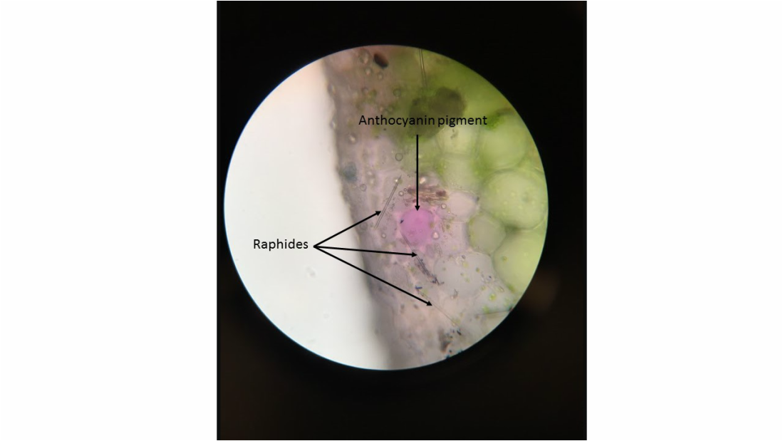
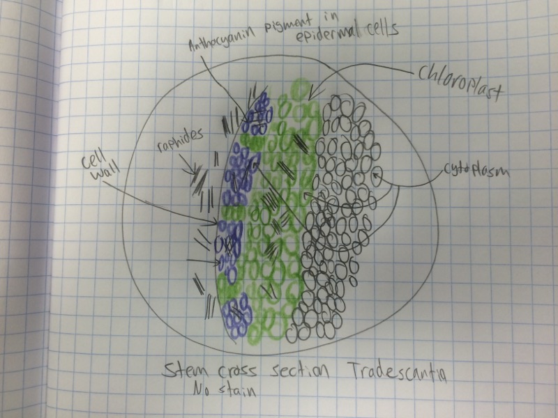
Anatomy of Tradescantia fluminensis ab Cross section of stem, cf Cross section of leaf, g Fragment of leaf adaxial epidermis, h Fragment of leaf abaxial. Anatomy of Tradescantia fluminensis ab Cross section of stem, cf Cross section of leaf, g Fragment of leaf adaxial epidermis, h Fragment of leaf abaxial. Stem crosssection of Tradescantia (Spiderwort) This is a stem crosssection of Tradescantia (Spiderwort) with no stain at a magnification of 100X At the very tip of the microscope's eyepiece pointer are a grouping of raphides (needlelike crystals made of calcium oxalate) that were exposed due to the rupturing of the vacuoles.
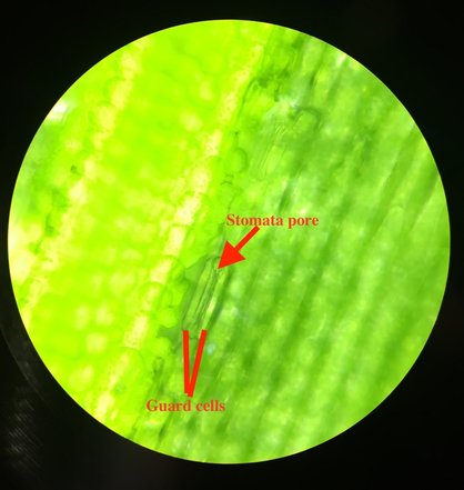
Download 1,309 Cross Section Leaf Stock Illustrations, Vectors & Clipart for FREE or amazingly low rates!. These cells can change shape in order to close the pore In very hot conditions water inside the leaf evaporates and the water vapour can escape through the stomata Closing them prevent reduces water loss, but also limits the diffusion of carbon dioxide and oxygen in and out of the leaf. Pine leaf Cross Section Link to detail of epidermis with stoma Link to detail of vascular tissue Return to the shoot page.
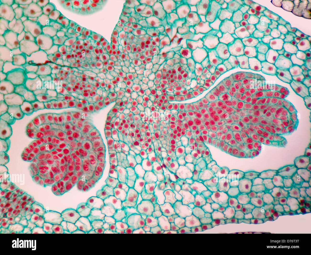
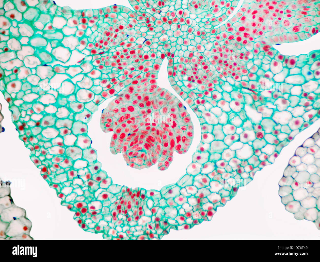
Fig 3 Cross section of the stem Ensemble (A, x 95) Portion with cortex and stele (B, x 175) Portion with vascular bundles (C, x 265) c cortex, cl chlorenchyma, e epidermis, gt ground tissue, ld lipid droplets, vb vascular bundle (the arrow indicates a calcium oxalate crystal) "Histoanatomical study on the vegetative organs of Tradescantia spathacea (Commelinaceae)". Elodea leaf cells with structures labeled Chloroplasts and mitochondria move within Elodea leaf cells;. One previous study was conducted, by Scott and Priestley (1925), on the anatomy 274 AÇÜ Orman Fak Derg (12) 13(2) vb li ss vb a 100µm t 100µm b ue st le c 100µm d 50µm ue vb bs le le cl e 50µm f 50µm st g 50µm h 50µm Figure 2 Anatomy of Tradescantia fluminensis a b Cross section of stem, c f Cross section of leaf, g.
10 _____ opening in leaf surface for gas exchange and water release 11 _____ organelle in plants where photososynthesis takes place Label these structures in a crosssection from a micrograph of a REAL leaf WORD BANK palisade mesophyll cuticle guard cells stoma epidermis. Mesophytic Leaf Anatomy Figure \(\PageIndex{4}\) Nerium leaf cross section Instead of sunken stomata, the epidermis in oleander recesses and creates a pocket that is lined with trichomes The stomata are located at the base of these pockets, called stomatal crypts The trichomes help capture evaporating moisture and maintain a relatively. Anatomy of Tradescantia fluminensis ab Cross section of stem, cf Cross section of leaf, g Fragment of leaf adaxial epidermis, h Fragment of leaf abaxial.
113 MB Picture Natural History No 316 Section of Leafpng 499 × 364;. Card 13 Prepare a drawing of a bean leaf cross section Label the tissues. Dicot leaf in cross section with branching veins II Internal (Microscopic) Anatomy of Monocot Leaves 1 On the same slide you used for the previous section, use the scanner objective to locate the cross section of the monocot leaf, then use greater magnification to find the following structural details Angiosperms 2.
Figure 2817 Leaf anatomy ;. A crosssection through a leaf Features of leaves and their functions Feature Function;. Pine leaf Cross Section Link to detail of epidermis with stoma Link to detail of vascular tissue Return to the shoot page.
Figure 2817a Leaf anatomy (part 1 cutaway drawing of leaf tissues) Figure 2817b Leaf anatomy (part 2 surface view of a leaf of a spiderwort, Tradescantia, LM) Figure 2817c Leaf anatomy (part 3 cross section of a leaf of a lilac, Syringa, LM) Figure 2818 Organization of primary tissues in young stems. This coloring sheet is comprised of two half sheets The first half sheet is a plant cell which is more common I needed it to be easier to color The other side of the sheet shows a cross section of a leaf The focus is the gas exchange from the stomata The arrows represent the different gases exc. Plant Tissues Chapter 28 * * Figure 286 Evolutionary adaptations of stems * * * * * Figure 2817b Leaf anatomy (part 2 surface view of a leaf of a spiderwort, Tradescantia, LM) * * Figure 2817a Leaf anatomy (part 1 cutaway drawing of leaf tissues) * Figure 2817c Leaf anatomy (part 3 cross section of a leaf of a lilac, Syringa, LM) * Figure 287 Evolutionary adaptations of leaves Tissue.
New users enjoy 60% OFF 153,909,424 stock photos online. Cross section of a leaf lamina diagram of cross section a leaf cross section of a leaf biology cross section of a leaf biology Draw A Labelled Diagram Of Cross Section Leaf Lamina To Show Chloroplasts From Science Life Processes Class 10 Cbse Draw The Diagram Of Cross Section A Leaf And Label Chloroplast Cuticle Brainly In. Tradescantia zebrina (spiderwort) Perform a cross and a paradermal section Perform a cross and a paradermal section The simplest case we could imagine for an epidermis would be like the upper (adaxial) epidermis on the Tradescantia leaf.
Diagram The fullsize printout is available only to site members cuticle the waxy, waterrepelling layer on the top and bottom surfaces of a leaf;. Illustration of Plant Tissue Systems vector illustration Labeled biological structure scheme Anatomical diagram with leaf, stem and root microscopic graphic Plant inner vascular, dermal and ground cross section vector art, clipart and stock vectors Image. Label the tissues Make freehand cross sections of a bean leaf by placing a piece of it between two blocks of carrot root Stain and mount your sections Using the compound microscope, identify the tissues Which type of tissue composes the leaf veins?.
Plant Anatomy DRAFT 5th 9th grade 299 times Biology 68% average accuracy 3 years ago jcteach 0 Save Edit Edit. The above is a cross section of a xerophytic (found in dry habitats) leaf Xerophytic leaves typically have a thick cuticle to minimize water loss through transpiration and enlarged parenchyma cells for water storage that lack chloroplasts (water cells). These cells can change shape in order to close the pore In very hot conditions water inside the leaf evaporates and the water vapour can escape through the stomata Closing them prevent reduces water loss, but also limits the diffusion of carbon dioxide and oxygen in and out of the leaf.
Valora esta carrera Plan de estudios;. Start studying Plant Science Images Learn vocabulary, terms, and more with flashcards, games, and other study tools. (a) Draw the diagram of cross section of a leaf and label the following parts (i) chloroplast (ii) cuticle (b) A gas is released during photosynthesis Name the gas and also state the way in which the gas is evolved (c) In certain group of plants, stomata remains closed during day How is food synthesized by such plants Also name them.
Nymphaea leaf crosssectionjpg 1,424 × 1,512;. 21/11/19 at 906 AM Reply Mesophyll is differentiated into palisade (has parallel arranged elongated cells) and parenchyma (with loosely arranged oval/round cells) Cuticle A waxy layer that prevent water loss by evaporation Depending on how it has been cut, the crosssection of a cylinder may be either circle, rectangle, or oval Cross Section Of A Leaf. Leaf blade crosssection the leaf blade is more or less flat in crosssection 2×Tradescantia subaspera KerGawl is a very rare spiderwort hybrid likely originating from a garden planting It is known from MA It has glandular hairs on the sepals.
Describes the structure and function of leaves We have moved all content for this concept to for better organization Please update your bookmarks accordingly. 10 _____ opening in leaf surface for gas exchange and water release 11 _____ organelle in plants where photososynthesis takes place Label these structures in a crosssection from a micrograph of a REAL leaf WORD BANK palisade mesophyll cuticle guard cells stoma epidermis. Tradescantia virginiana NC State University and NC A&T State University work in tandem, along with federal, state and local governments, to form a strategic partnership called NC Cooperative Extension, which staffs local offices in all 100 counties and with the Eastern Band of Cherokee Indians.
2 Examine the prepared slide of a cross section through a leaf under the compound microscope Draw a neat, clear diagram of your specimen in the space below Find all of the structures illustrated above and label them 3 Obtain a specimen of a Tradescantia leaf from your teacher Place a flat section of the leaf bottomside up on a slide as a. It helps keep the leaf from dying out (and protects it from invading bacteria, insects, and. Leaf Anatomy Mini Lab (15 points) Comparing three types of plant cells under the microscope Name Role Arielle Zappia Microscopic work Michael Lee Scribe (and research) data table, magnification, labeling cell Jacob Zhang Research (and scribe) how do the plant’s different environments affect how they look under the microscope?.
Card 13 Prepare a drawing of a bean leaf cross section Label the tissues. Leaf Cross Section Under the Microscope Whereas the transparent thin epidermal skin of the leaf allows the student to observe the stomata and other epidermal cells, it would be important to prepare a cross section of a leaf to observe the arrange of cells inside the leaf structure Requirements. 371 KB Pter1026 fern leaf Ljpg 1,0 × 736;.
Label the tissues Make freehand cross sections of a bean leaf by placing a piece of it between two blocks of carrot root Stain and mount your sections Using the compound microscope, identify the tissues Which type of tissue composes the leaf veins?. Mesophytic Leaf Anatomy Figure \(\PageIndex{4}\) Nerium leaf cross section Instead of sunken stomata, the epidermis in oleander recesses and creates a pocket that is lined with trichomes The stomata are located at the base of these pockets, called stomatal crypts The trichomes help capture evaporating moisture and maintain a relatively. This Leaf Cross Section Color Unlabeled clipart is great to illustrate your teaching materials As an abcteach member you have unlimited access to our 22,000 clipart illustrations and can use them for commercial use This Leaf Cross Section Color Unlabeled clipart is provided in jpeg format.
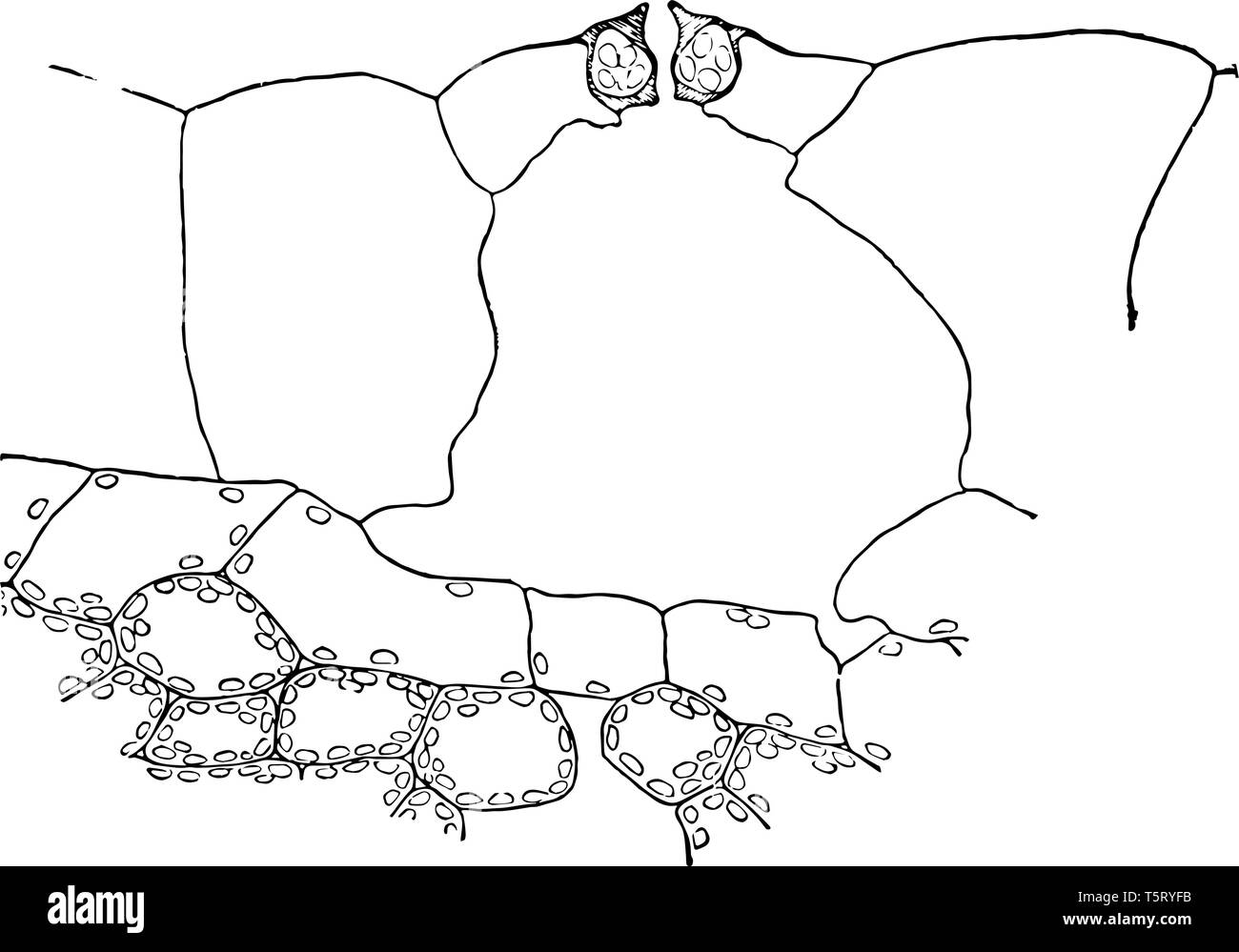
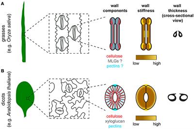
Spectrum that corresponds well to our experimentally found spectra We use SEM on crosssections of Tradescantia leaves to estimate the local variation in cell wall thickness (Figure S8) Fig S8 SEM images of a cross section of the adaxial side of a Tradescantia leaf In each image the thickness of the cell wall is indicated, showing large. Cross section of a leaf lamina diagram of cross section a leaf cross section of a leaf biology cross section of a leaf biology Draw A Labelled Diagram Of Cross Section Leaf Lamina To Show Chloroplasts From Science Life Processes Class 10 Cbse Draw The Diagram Of Cross Section A Leaf And Label Chloroplast Cuticle Brainly In. Evelyn Bailey Leaf vascular tissue is located within the mesophyll layer Model 2 — Cross Section of the Internal Structure of a Leaf Cuticle Upper epidermis Chloroplast palisade mesophyll Air Space Lower epidermis Vein Spongy mesophyll Cuticle Guard cell f Stoma 8 You can see these if you look at a transverse section (crosssection) of a leaf under a microscope Bundlesheath cells.
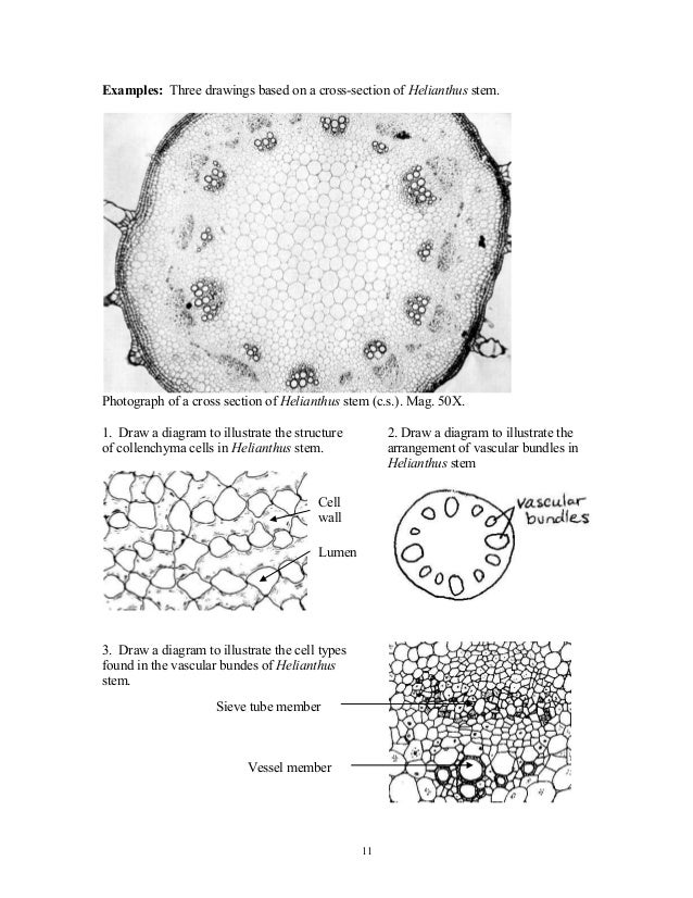
1 Select Cross Sections > Update Cross Section from the InRoads bar menu 2 On the General leaf, select the Cross Section Set to be updated using the drop down menu or the “target” button then on the desired cross section 3 Toggle on the desired Mode These are ♦ Refresh used to update existing cross section data. Nuclei are also visible as clear, friedeggshaped structures Elodea are common freshwater aquarium plants An elodea leaf was mounted in pondwater between a slide and coverslip with a silicon spacer Images were taken on an inverted. Crosssection of a stem of corn (Zea mays) 5 Organization of Stems Woody Eudicot Stem Stem Crosssection and Wood Blocks 1) pp (top) Read the descriptions of terms and using the labeled photos of the stem crosssections (Fig 97), identify the tisssues in a prepared slide of a crosssection of a 3 yr old stem of a basswood tree.
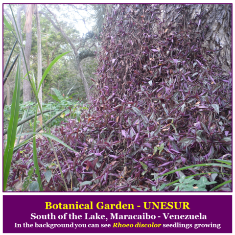
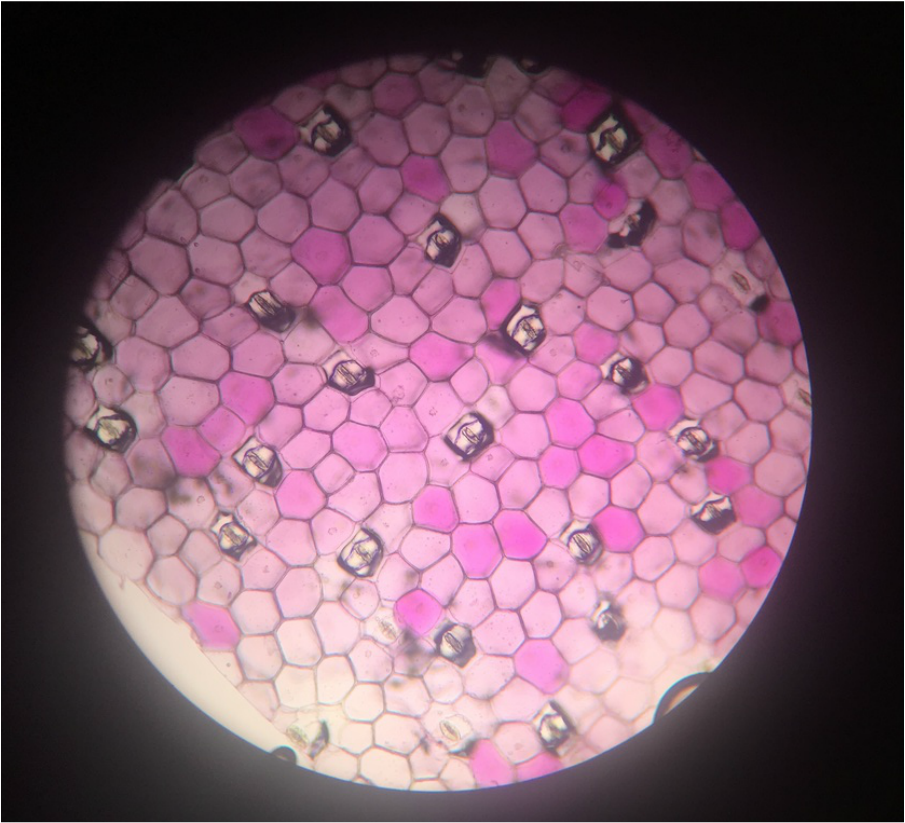
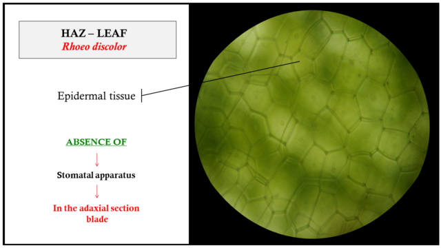
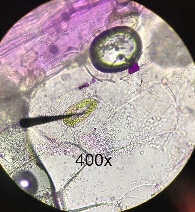
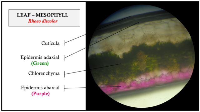
Description Tradescantia spathacea is a rather succulent small and lowgrowing plant, with rosettes of waxy lanceshaped leaves metallic green above, with glossy purple underneath This plant is also known as Rhoeo discolor and more frequently in the horticulture trade as Rhoeo spathaceaThe species name is a Latin word referring to the spathelike large bracts which envelope the flower. Tradescantia upper leaf epidermis x100 Note the lack of stomata on this leaf surface The black dots are nuclei Unlike this species, many dicot plants have a few stomata on their upper surface Most of a dicot leaf's stomata are usually found on the lower surface Tradescantia lower leaf epidermis x100 There are lots of pairs of guard cells. (Tradescantia) leaf (LM) Epidermal cell (b) m Fig 3518c Upper epidermis Palisade mesophyll Key to labels Dermal Ground Vascular Spongy mesophyll Lower epidermis Vein Air spaces Guard cells Cross section of a lilac (Syringa) leaf (LM) (c) µm Secondary Growth •Secondary growth adds girth to stems and roots in woody plants, (it doesn’t.

Tradescantia An Overview Sciencedirect Topics
Plant Stomata Under The Microscope And What Stomata Tell You About Plant Habitat

Leaves The Ohio State University At Lima
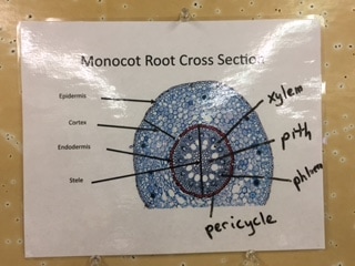
Monocot Root Cross Section Of A Corn Plant Osu Plant Structure Bot313 Winter 17
Www Bxscience Edu Ourpages Auto 11 3 7 Activity 24 plant Structure slides Pdf

Pdf Contributions To The Leaf And Stem Anatomy Of Tradescantia Fluminensis An Alien Species New To The Flora Of Turkey

Pdf Contributions To The Leaf And Stem Anatomy Of Tradescantia Fluminensis An Alien Species New To The Flora Of Turkey

Chapter 32 Leaf Structure And Function Ppt Video Online Download

Plant Science Images Flashcards Quizlet

Anatomy Of Tradescantia Fluminensis A B Cross Section Of Stem C F Download Scientific Diagram
Parts Of The Cell Class 8 Biology

Unbiol1
Page 2 Cell Membrane Cross Section High Resolution Stock Photography And Images Alamy

Pin On Voronoi
Category Stomata Osu Plant Structure Bot313 Winter 17

Leaf Stoma Tradescantia Spathacea Under Microscopy Stock Photo Edit Now
Tradescantia Wikipedia
Category Vascular Bundles Osu Plant Structure Bot313 Winter 17
Hetal Craig Armstrong Plant Biology Lab
Stoma Plant High Resolution Stock Photography And Images Alamy
Q Tbn And9gcqb172vxrfsx0p13izjl3uiuq1n5ddk Dxsclr69l2sejtsivt Usqp Cau
Athena Kelsey Armstrong Plant Biology Lab

Anatomy Of Tradescantia Fluminensis A B Cross Section Of Stem C F Download Scientific Diagram
Frontiers Balancing Strength And Flexibility How The Synthesis Organization And Modification Of Guard Cell Walls Govern Stomatal Development And Dynamics Plant Science
Leaf Cross Section Diagram Label Worksheets Differentiated Teaching Resources
Www Bxscience Edu Ourpages Auto 11 3 7 Activity 24 plant Structure slides Pdf
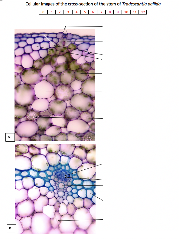
Solved Cellular Images Of The Cross Section Of The Stem O Chegg Com

Chapter 28 Plant Structure And Growth Ppt Download
Bio311
Blog Posts Osu Plant Structure Bot313 Winter 17
Www Nzqa Govt Nz Assets Qualifications And Standards Qualifications Ncea Ncea Subject Resources Biology Exp Pdf
Q Tbn And9gcqgp0b Sidx3zvmlaqgdm4du0ittsmfcmdxc Sp6j7obkvspa9g Usqp Cau
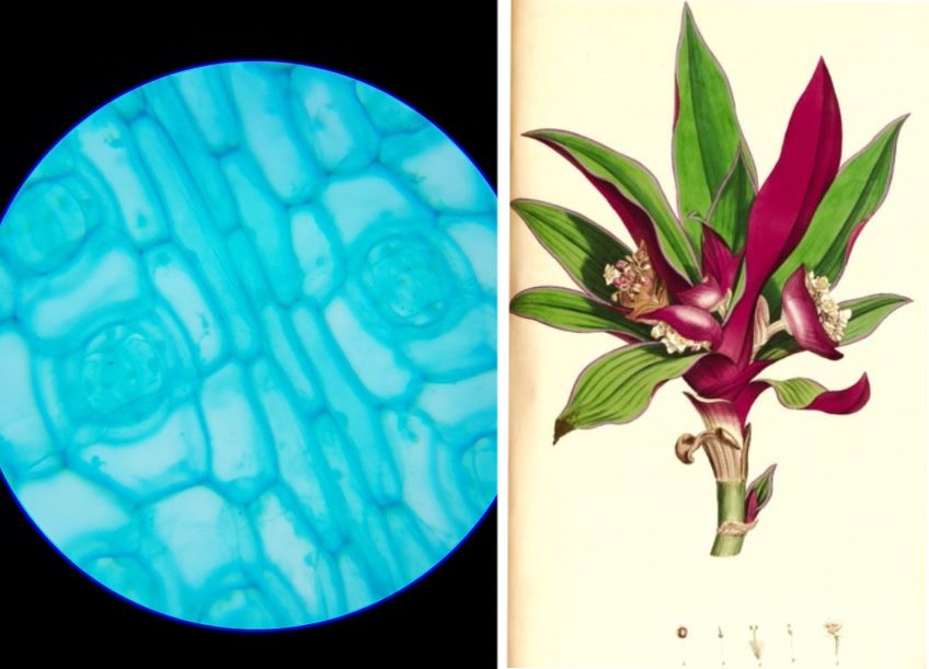
Rhoeo Discolor Leaf W M Under The Microscope Steemit

Copyright C 05 Pearson Education Inc Publishing As Benjamin Cummings Chapter 35 Plant Growth And Development Ppt Download
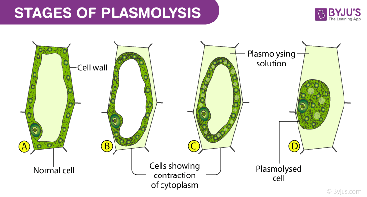
Study Of Plasmolysis In Epidermal Peels Example Rhoeo Leaves
Page 2 Cell Membrane Cross Section High Resolution Stock Photography And Images Alamy

Chapter 35 Presentation

Leaf Structure And Function Read Biology Ck 12 Foundation
Organography And Plant Histology Of Rhoeo Discolor Tradescantia Spathacea Steemit
Www Gtac Edu Au Wp Content Uploads 16 01 Leaf Epidermal Peel Labpreparation Pdf
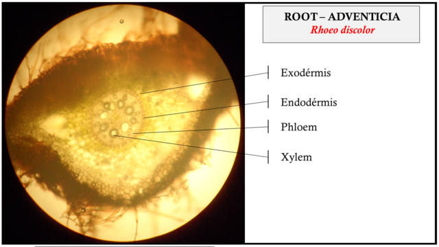
Organography And Plant Histology Of Rhoeo Discolor Tradescantia Spathacea Steemit

Unbiol1

Stoma Wikipedia

Leaves The Ohio State University At Lima

Unbiol1

Leaves The Ohio State University At Lima
Page 2 Cell Membrane Cross Section High Resolution Stock Photography And Images Alamy

Organography And Plant Histology Of Rhoeo Discolor Tradescantia Spathacea Steemit

Leaves The Ohio State University At Lima
Bio311

Pdf Contributions To The Leaf And Stem Anatomy Of Tradescantia Fluminensis An Alien Species New To The Flora Of Turkey Ozgur Eminagaoglu Academia Edu
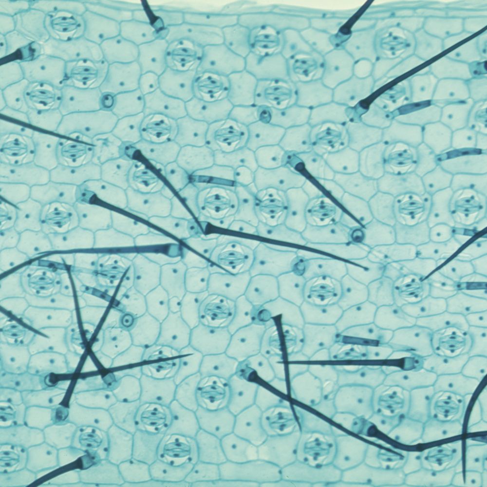
Spiderwort Leaf Epidermis W M Microscope Slide Carolina Com

Leaves The Ohio State University At Lima

Monocot Leaf Cross Section 0x Plant Structure Cross Section Plant Pathology

Josh Brendin Armstrong Plant Biology Lab

Leaves The Ohio State University At Lima

Leaves The Ohio State University At Lima
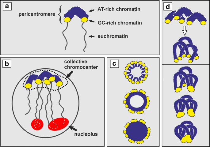
Pericentromere Clustering In Tradescantia Section Rhoeo Involves Self Associations Of At And Gc Rich Heterochromatin Fractions Is Developmentally Regulated And Increases During Differentiation Springerlink
Lab 2 Test Flashcards Chegg Com

Anatomy Of Tradescantia Fluminensis A B Cross Section Of Stem C F Download Scientific Diagram

Chapter 35 Presentation

Tradescantia Wikipedia

Shortroot Mediated Increase In Stomatal Density Has No Impact On Photosynthetic Efficiency Plant Physiology

Chapter 32 Leaf Structure And Function Ppt Video Online Download

Cbse Papers Questions Answers Mcq Cbse Class 8 Ch8 Science Cell Structure And Functions Mcqs
Category Corn Osu Plant Structure Bot313 Winter 17
Athena Kelsey Armstrong Plant Biology Lab

Leaves The Ohio State University At Lima
Category Vascular Bundles Osu Plant Structure Bot313 Winter 17
Plant Structure Growth And Development Ppt Download
Www Bxscience Edu Ourpages Auto 11 3 7 Activity 24 plant Structure slides Pdf

Anatomy Of Tradescantia Fluminensis A B Cross Section Of Stem C F Download Scientific Diagram
Q Tbn And9gcqsxbca4su2esbwjbgkbziskc2uray3nydrk7kzgtxkhapt7etz Usqp Cau

Tradescantia An Overview Sciencedirect Topics

Unbiol1

Unbiol1

Monocots Vs Eudicots Plant Structure Bot315

Tradescantia Wikipedia

Epidermis Peel Cell Wall Microscopic Cells Cell
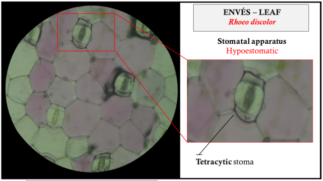
Organography And Plant Histology Of Rhoeo Discolor Tradescantia Spathacea Steemit

Structure And Function Of Cells Learn Biology Class 8 Amrita Vidyalayam Elearning Network

Amazing Photos Of Microscopic Subjects From The 14 Nikon Small World Competition Nikon Small World Microscopic Photography Mind Blowing Images

Chapter 35 Plant Structure Growth And Development Ppt Video Online Download
Category Stomata Osu Plant Structure Bot313 Winter 17

Monocotyledon An Overview Sciencedirect Topics

Leaves The Ohio State University At Lima
Leaves
Http Biozoojournals Ro Bihbiol Cont V3n1 Chimpan Pdf
Q Tbn And9gcqgp0b Sidx3zvmlaqgdm4du0ittsmfcmdxc Sp6j7obkvspa9g Usqp Cau
Hetal Craig Armstrong Plant Biology Lab
Bio311
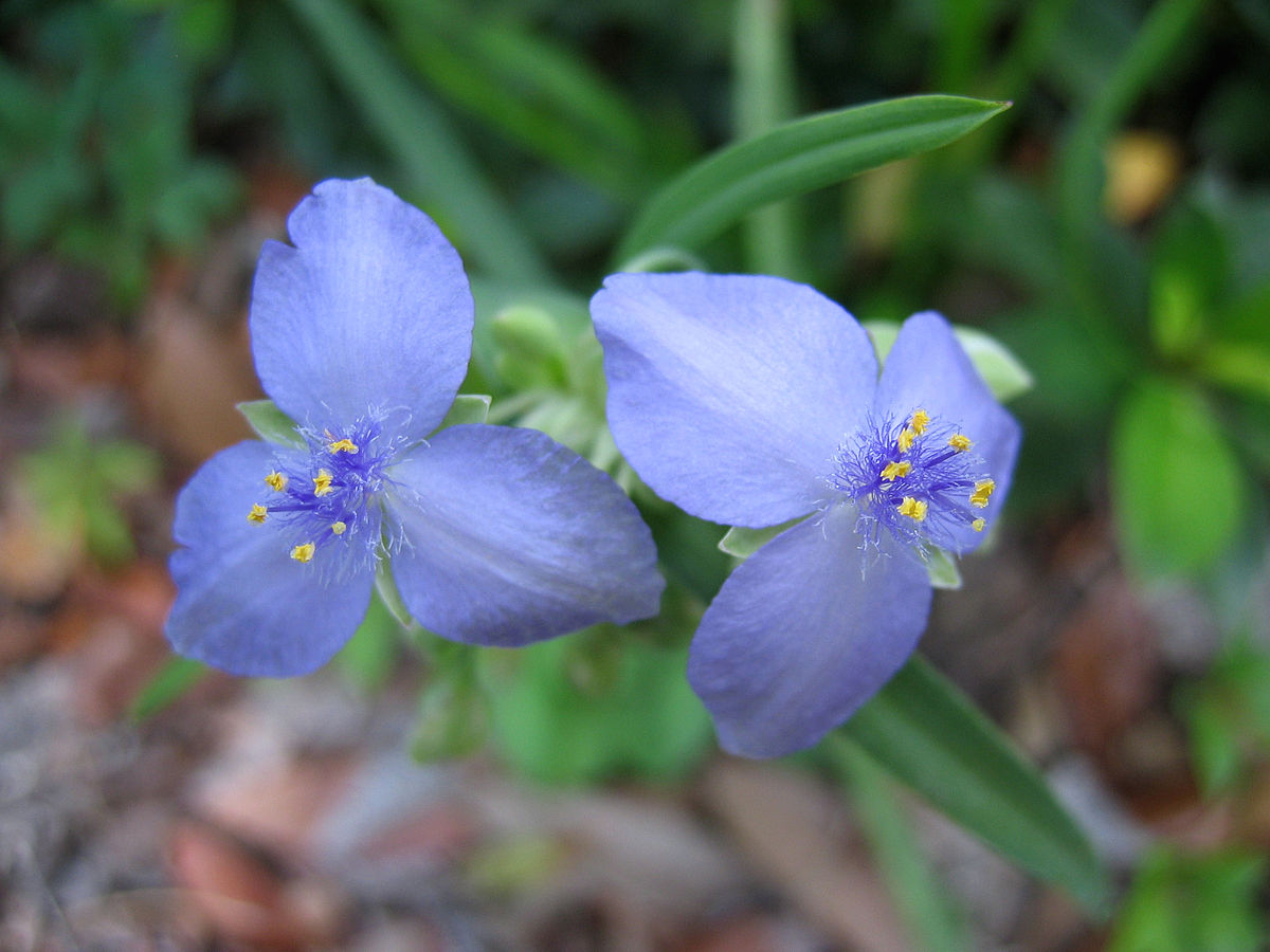
Tradescantia Wikipedia
Kevin And Loyes Great Plant Adventures Armstrong Plant Biology Lab
.PNG)
The Vascular Cambium And Secondary Vascular Tissue

Leaf Stoma Tradescantia Spathacea Under Microscopy Stock Photo Edit Now

Leaves The Ohio State University At Lima

Tradescantia
Organography And Plant Histology Of Rhoeo Discolor Tradescantia Spathacea Steemit


